45 microscope labeled diagram
Microscope Types (with labeled diagrams) and Functions Simple microscope labeled diagram Simple microscope functions It is used in industrial applications like: Watchmakers to assemble watches Cloth industry to count the number of threads or fibers in a cloth Jewelers to examine the finer parts of jewelry Miniature artists to examine and build their work Also used to inspect finer details on products Microscope labeled diagram - SlideShare Microscope labeled diagram 1. The Microscope Image courtesy of: Microscopehelp.com Basic rules to using the microscope 1. You should always carry a microscope with two hands, one on the arm and the other under the base. 2. You should always start on the lowest power objective lens and should always leave the microscope on the low power lens ...
Labeled Diagram Of A Stereo Microscope - GlobalSpec Products/Services for Labeled Diagram Of A Stereo Microscope. Microscopes - (705 companies) Microscopes are instruments that produce magnified images of small objects Microscopes are instruments that produce a magnified image of a small object. They are used in many scientific and industrial applications.

Microscope labeled diagram
Animal Cell Diagram Under Light Microscope Labeled Tuesday, April 20th 2021. | Diagram. Animal Cell Diagram Under Light Microscope. To make observations and draw scale. This shows a generalized animal cell under a light microscope. We all keep in mind that the human physique is amazingly elaborate and one way I discovered to comprehend it is by way of the style of human anatomy diagrams. Parts of the Microscope with Labeling (also Free Printouts) Parts of the Microscope with Labeling (also Free Printouts) A microscope is one of the invaluable tools in the laboratory setting. It is used to observe things that cannot be seen by the naked eye. Table of Contents 1. Eyepiece 2. Body tube/Head 3. Turret/Nose piece 4. Objective lenses 5. Knobs (fine and coarse) 6. Stage and stage clips 7. Aperture Microscope Diagram and Quiz | Science diagrams, Science printables ... Description Worksheet identifying the parts of the compound light microscope. Answer key: 1. Body tube 2. Revolving nosepiece 3. Low power objective 4. Medium power objective 5. High power objective 6. Stage clips 7. Diaphragm 8. Light source 9. Eyepiece 10. Arm 11. Stage 12. Coarse adjustment knob 13. Fine adjustment knob 14. Base S
Microscope labeled diagram. Microscope Parts, Function, & Labeled Diagram - slidingmotion Microscope parts labeled diagram gives us all the information about its parts and their position in the microscope. Microscope Parts Labeled Diagram The principle of the Microscope gives you an exact reason to use it. It works on the 3 principles. Magnification Resolving Power Numerical Aperture. Parts of Microscope Head Base Arm Eyepiece Lens Microscope Parts Diagram Quiz - PurposeGames.com This is an online quiz called Microscope Parts Diagram. There is a printable worksheet available for download here so you can take the quiz with pen and paper. Your Skills & Rank. Total Points. 0. Get started! Today's Rank--0. Today 's Points. One of us! Game Points. 15. You need to get 100% to score the 15 points available. Labeling the Parts of the Microscope Labeling the Parts of the Microscope. This activity has been designed for use in homes and schools. Each microscope layout (both blank and the version with answers) are available as PDF downloads. You can view a more in-depth review of each part of the microscope here. Compound Microscope Labeled Diagram | Quizlet QUESTION. The total magnification of a specimen being viewed with a 10X ocular lens and a 40X objective lens is. 15 answers. QUESTION. a mosquito beats its wings up and down 600 times per second, which you hear as a very annoying 600 Hz sound. if the air outside is 20 C, how far would a sound wave travel between wing beats. 2 answers.
Parts of a microscope with functions and labeled diagram Figure: Diagram of parts of a microscope There are three structural parts of the microscope i.e. head, base, and arm. Head - This is also known as the body. It carries the optical parts in the upper part of the microscope. Base - It acts as microscopes support. It also carries microscopic illuminators. Compound Microscope Parts - Labeled Diagram and their Functions - Rs ... Labeled diagram of a compound microscope Major structural parts of a compound microscope There are three major structural parts of a compound microscope. The head includes the upper part of the microscope, which houses the most critical optical components, and the eyepiece tube of the microscope. Parts of Stereo Microscope (Dissecting microscope) - labeled diagram ... Labeled part diagram of a stereo microscope Major structural parts of a stereo microscope There are three major structural parts of a stereo microscope. The viewing Head includes the upper part of the microscope, which houses the most critical optical components, including the eyepiece, objective lens, and light source of the microscope. PDF Parts of a Microscope Printables - Homeschool Creations Label the parts of the microscope. You can use the word bank below to fill in the blanks or cut and paste the words at the bottom. Microscope Created by Jolanthe @ HomeschoolCreations.net. Parts of a eyepiece arm stageclips nosepiece focusing knobs illuminator stage objective lenses
Labelled Diagram of Compound Microscope - Biology Discussion The below mentioned article provides a labelled diagram of compound microscope. Part # 1. The Stand: The stand is made up of a heavy foot which carries a curved inclinable limb or arm bearing the body tube. The foot is generally horse shoe-shaped structure (Fig. 2) which rests on table top or any other surface on which the microscope in kept. (a) Draw the labelled ray diagram for the formation of image by a ... Click here👆to get an answer to your question ️ (a) Draw the labelled ray diagram for the formation of image by a compound microscope. Derive an expression for its total magnification (or magnifying power), when the final image is formed at the near point.(b) Why both objective and eyepiece of a compound microscope must have short focal lengths?Draw a ray diagram showing the image ... Microscope Anatomy Diagram | Quizlet This test will help the student to identify the different parts and function of the microscope. Terms in this set (12) Arm Supports the tube and connects it to the base Eyepiece the lens at the top that you look through. They are usually 10X or 15X power. Base The bottom of the microscope, used for support Body tube Parathyroid Gland Histology with Microscope Slide Image and Labeled Diagram Parathyroid Gland Histology with Microscope Slide Image and Labeled Diagram 09/12/2021 07/12/2021 by anatomylearner There are two or more pairs of parathyroid glands located on the posterior surface of the thyroid gland. You will find two main types of cells (chief and oxyphils) in the parathyroid gland histology slide.

Under the Micrsocope: Onion Cell (100x - 400x) - YouTube
Animal Cell Diagram Under Microscope Labeled Draw a large diagram of an animal cell as seen through an electron microscope. Label the parts … (Myra Burns) If you want to download the image of Plant Cell Worksheet together with Structure Of Animal Cell and Plant Cell Under Microscope Diagrams, simply right click the image and choose "Save As".

Human Blood under microscope - YouTube
Neuron under Microscope with Labeled Diagram » AnatomyLearner >> The ... Let's see the neuron histology slide labelled diagram and try to find out the below-mentioned characteristics - Presence of an identifiable cell body (soma) that locates in the brain's grey matter (according to the slide image). The cell body possesses spherical, euchromatic, and large eccentric nuclei containing a prominent nucleolus.
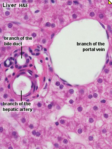
Male Reproductive System | histology
PDF Label parts of the Microscope: Answers Label parts of the Microscope: Answers Coarse Focus Fine Focus Eyepiece Arm Rack Stop Stage Clip . Created Date: 20150715115425Z ...
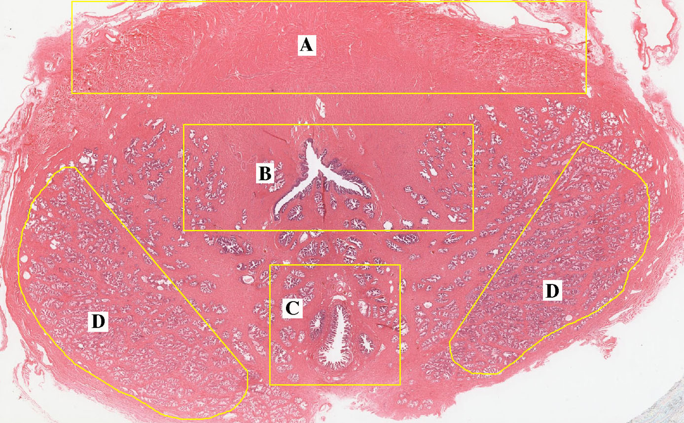
ANAT2511 Gastrointestinal Tract - Embryology
Compound Microscope Parts, Functions, and Labeled Diagram Compound Microscope Parts, Functions, and Labeled Diagram Posted by Fred Koenig on Nov 18th 2020 Compound Microscope Parts, Functions, and Labeled Diagram Parts of a Compound Microscope Each part of the compound microscope serves its own unique function, with each being important to the function of the scope as a whole.

Chloroplasts moving by cytoplasmic streaming in the cells of the ...
Microscope, Microscope Parts, Labeled Diagram, and Functions 19 Jan 2022 — The Microscopes parts divided into three different structural parts Head, Base, and Arms. ... Head/Body: It contain the optical parts in the upper ...Microscope Parts: Microscope Parts FunctionsObjective lenses: Low-, medium-, and high-po...Light source: Provides light for viewing the spe...Base: Supports the microscope
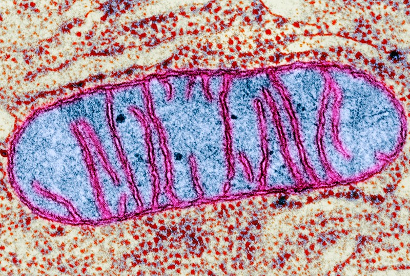
Mitochondrial genome editing: another win for curiosity-driven research
Label the microscope — Science Learning Hub All microscopes share features in common. In this interactive, you can label the different parts of a microscope. Use this with the Microscope parts activity to help students identify and label the main parts of a microscope and then describe their functions. Drag and drop the text labels onto the microscope diagram.

Neurolemmocyte On Skeletal Muscle Model - Human Anatomy - GUWS Medical
Inverted Microscope- Definition, Principle, Parts, Labeled Diagram ... This is a reverse of the normal construction of a microscope, where the objective lenses are found above the stage while the condenser and the light source are below the stage. Hence the word, 'inverted'. And therefore, instead of viewing the image from up, downwards, with the inverted microscope you view the image from down, upwards.
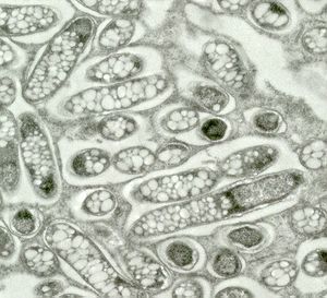
Bradyrhizobium - microbewiki
Microscope Parts and Functions With Labeled Diagram and Functions How ... Most specimens are mounted on slides, flat rectangles of thin glass. The specimen is placed on the glass and a cover slip is placed over the specimen. This allows the slide to be easily inserted or removed from the microscope. It also allows the specimen to be labeled, transported, and stored without damage.
Post a Comment for "45 microscope labeled diagram"