43 label the internal structures of the clam
PDF Taxonomy, Anatomy, and Biology of the Hard Clam Internal Clam 1 Mantle Shell Anatomy • Covers visceral or body mass • Holds in fluid • Secrets new shell 2. Ant. adductor muscle 3. Post adductor musclePost. adductor muscle • Hold valves shut 4. Pericardium cavity • Region covered with thin Region covered with thin, dark membrane • Contains 2-chambered heart and kidney in a fluid-filled sac 5. Clam Dissection - JKL Bahweting Middle School Label the internal structures of the clam and draw red arrows showing the pathway of food as it travels to the clam's stomach: Click on image to enlarge Guided Exploration 1. What is the oldest part of a clam's shell called and how can it be located? 2. What do the rings on the clam's shell indicate? 3. Name the clam's siphons. 4.
Clam Lab Key Terms Questions and Study Guide - Quizlet Guides food into mouth (clam). Gills: Extensions of the body containing thin-walled blood vessels that allow for easy absorption of oxygen from the outside surface. Digestive Glands: What an organ secretes enzymes into the pyloric chamber of the stomach to assist in the chemical digestion of food. Visceral Mass:

Label the internal structures of the clam
PDF Biology 11 Name: Clam Dissection 1 anterior posterior 2. - Mrs Dildy Clam Dissection Purpose: To examine the internal structure of a bivalve Procedure: 1. Observe the external anatomy of the clam, identify the anterior and posterior ends of the clam as well as the dorsal, ventral, & lateral surfaces. 2. Locate the umbo, the bump at the anterior end of the valve. This is the oldest part of the clam shell. Clam Dissection Questions - vmsteacher.org 6. Where is the mantle located in the clam? What is its function? 7. How do clams breathe? 8. What helps direct water over the gills? 9. Where are the palps found and what is their function? 10. Where is the clam's heart located? 11. Label the internal structures of the clam and draw arrows showing the pathway of food as it travels to the clam ... DOC Clam Dissection - js082.k12.sd.us The tube-like structure that runs through the pericardium is the intestine. Date: _____ Name: _____ 25. Answer the questions on your lab report & label the diagrams of the internal structures of the clam. Also, use arrows on the clam diagram to trace the pathway of food as it travels to the clam's stomach.
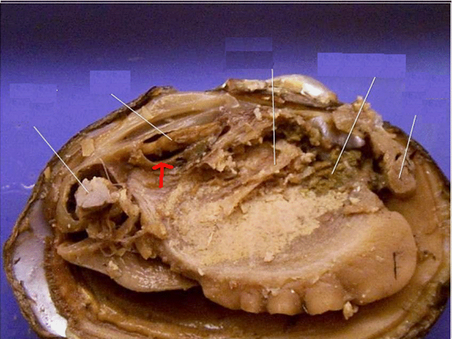
Label the internal structures of the clam. Solved Results 1. Draw a diagram and label the internal | Chegg.com Draw a diagram and label the internal anatomy of the clam. Which structures were difficult to locate? 2. In what ways does the clam differ from other mollusks? What modifications are critical for its unique habitat and lifestyle? Question: Results 1. Draw a diagram and label the internal anatomy of the clam. Which structures were difficult to ... PDF CLAM DISSECTION HANDOUT - Cathy Ramos Expose the clam's interior anatomy by gently separating the mantle from the upper valve with a blunt probe and then lifting the upper valve. 4. Lift and fold the top mantle to expose the gills and body cavity. Find the body structures listed below. Draw and label the internal parts of your clam. See Table 6—1 for definitions of the parts. a ... Clam Dissection Questions - BIOLOGY JUNCTION What are the parts of the clam's nervous system? 17. Why are clam's referred to as "filter feeders"? 18. Label the internal structures of the clam and draw arrows showing the pathway of food as it travels to the clam's stomach: BACK Biology Junction Team April 21, 2017 Invertebrate Unit, My Classroom Material Clam Dissection - BIOLOGY JUNCTION Locate the muscle "scars" on the inner surface of the left valve. The adductor muscleswere attached here to hold the clam closed. Identify the mantle, the tissue that lines both valves & covers the soft body of the clam. Find the mantle cavity, the space inside the mantle. Locate two openings on the posterior end of the clam.
Clam Dissection.docx - Google Docs Place a clam in a dissecting tray and identify the anterior and posterior ends of the clam as well as the dorsal, ventral, & lateral surfaces. Figure 1 Figure 1 Locate the umbo, the bump at the... Bivalve Anatomy - Paleontological Research Institution The soft body inside of the shell includes a muscular foot, gills (or ctenidia, used for respiration and feeding), muscles, a digestive system, nerves, a three-chambered heart and an open circulatory system (with sinuses). Some species, such as the Hard-Shelled Clam here, have siphons for directing water flow in and out of the body chamber. What are parts of the clams nervous system? - FindAnyAnswer.com The labial palps gather the food and place it into the clam's mouth. The nervous system of clams consists of three pairs of ganglia connected by nerve cords. ... which are connected by a hinge joint and a ligament that can be external or internal. Clams also have kidneys, a heart, a mouth, a stomach, and a nervous system. Many have a siphon ... PDF Label the Clam Anatomy @Sheri Amsel Label the Clam Anatomy @Sheri Amsel . Created Date: 6/8/2018 3:46:07 PM
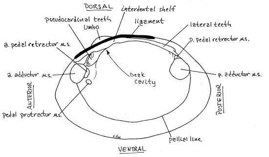
PDF Clam Anatomy Exercise Inside of the clam shell (use shucked clam): 1) Identify anterior and posterior ends, and the dorsal and ventral sides of clam shell below. 2) Locate the following on the clam shell below and record on blank lines provided: anterior and posterior muscle scars, umbo, hinge ligament and teeth, mantle attachment, and small teeth 8 where is the mantle located in the clam what is its Label the internal structures of the clam and draw arrows showing the pathwayof food as it travels to the clam's stomach: ANUSPosterior Adductor Muscle Excurrent Siphon Incurrent Siphon GILLSUMBO Intestine SHELL / Valve MANTLEFOOTHEART STOMACH MOUTH Anterior Adductor Muscle Labial Palp Intestine GONAD Clam Diagram Labeled - schematron.org Sep 25, 2018 · Structures to pin and label: 1. excurrent siphon, 2. incurrent siphon, 3. valve, 4. foot, 5. umbo, 6. heart, 7. posterior adductor muscle, . Clam Anatomy Labeling Page. to show answer keys or other teacher-only items. Link to More Info About this Animal (with Labeled Body Diagram). Click Here. Study 25 Terms | clam dissection functions Flashcards | Quizlet (interior) these are the respiratory structures obtaining oxygen and getting food Foot (interior) A clam uses its singular foot to dig down into the sand and draw nourishment into its palps, movement Visceral Mass (interior) soft tissue above the foot that holds the clams body organs Palps
32 Label The Internal Structures Of The Clam - Labels 2021
Clam structures and functions Flashcards | Quizlet clam. clam dissection: internal anatomy: parts & functio…. Parts and functions of the starfish, clam, and cra…. Describe the transit of breast milk from lactocytes to nipple pores. The water of a swimming pool is hypotonic to our cells.
31 Label The Internal Structures Of The Clam - Labels Database 2020
DOC Clam Dissection - Wallingford Public Schools Continue following the intestine toward the posterior end of the clam. Find the . anus. just behind the posterior adductor muscle. Use your probe to trace the path of food & wastes from the incurrent siphon through the clam to the excurrent siphon. Answer the questions on your lab report & label the diagrams of the internal structures of the clam.
Solved: 1) Draw The External Features Of A Freshwater Muss... | Chegg.com
PDF Lab 5: Phylum Mollusca - Amherst 2. Study the figures of the internal structure of the clam. Locate the adductor and retractor muscles. The adductor muscles (which were cut in two to open the shell) close the valves, whereas the retractor muscles pull in the foot. Notice the large mass of the two adductor muscles, which allow for the prolonged closure of the valves.

bioweb images
Basic Clam Anatomy (Internal) Quiz - PurposeGames.com This is an online quiz called Basic Clam Anatomy (Internal) There is a printable worksheet available for download here so you can take the quiz with pen and paper. From the quiz author Try to label these parts of a clam/mollusk This quiz has tags. Click on the tags below to find other quizzes on the same subject. Anatomy clam internal mollusca
Clam Internal Anatomy - Anatomy Drawing Diagram
DOC Clam Dissection - PC\|MAC The tube-like structure that runs through the pericardium is the intestine. Date: _____ Name: _____ 25. Answer the questions on your lab report & label the diagrams of the internal structures of the clam. Also, use arrows on the clam diagram to trace the pathway of food as it travels to the clam's stomach.

Clam Dissection
How can you label the internal structure of a clam? - Answers Apr 29, 2014 · An Internal Structure is the way an organism looks on the outside and an External Structure is the looks on the outside. When you open the clam will all the internal organs be visible? No, they ...

Clam Dissection Questions
PDF Clam Dissection Guideline - Monadnock Regional High School clam shell is the umbo, and it is from the hinge area that the clam extends as it grows. I. Purpose: The purpose of this lab is to identify the internal and external structures of a mollusk by dissecting a clam. II. Materials: 2 pairs of safety goggles 2 pairs of gloves 1 preserved clam 1 dissecting tray 1 paper towel 1 pair of scissors
Clam Dissection
Clam dissection: A first step into dissection and anatomy for young ... This is a long thin strand of muscle that is often wavy and the part of the clam that you can see when the shell is slightly open. This is known as the dorsal body, and it is like a robe that covers the internal organs of the clam. Over time, the mantle secretes what will become the shell, allowing the animal to grow a larger home. Foot
Clam Dissection Lab Questions - MATES-Biology-I
Clam Dissection - js082.k12.sd.us Why are clams called bivalves? Procedure 1. Put on your lab apron & safety glasses. 2. Place a clam in a dissecting tray and identify the anterior and posterior ends of the clam as well as the dorsal, ventral, & lateral surfaces. Figure 1 Figure 1 The left valve is on top if your clam is correctly positioned. The siphons are at the posterior end.
Post a Comment for "43 label the internal structures of the clam"