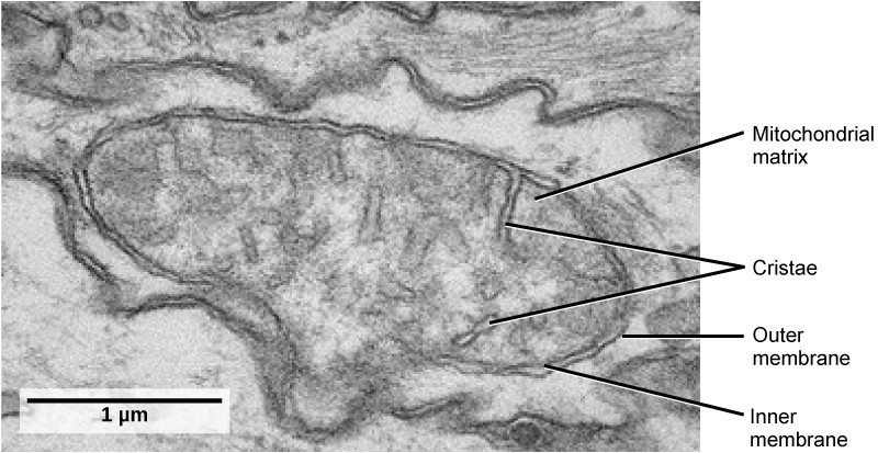
45 label the transmission electron micrograph of the mitochondrion.
Dynamics of mitochondrial cristae. (a) Transmission electron microscopy ... The use of an MTS for targeted delivery of the desired proteins to the mitochondria is widely applied to deliver FPs for mitochondria labeling or to influence mitochondrial function (Hoffmann et ... Transmission Electron Microscopy for Analysis of Mitochondria in Mouse ... May 20, 2018 · Transmission electron microscopy (TEM) is a powerful technique for ultrastructural studies ( Watson, 1958 ). TEM has been very useful in studying mitochondrial structure in skeletal muscle in both physiological and pathological conditions ( Picard et al., 2013 ).
Transmission electron microscopic images of chloroplasts ... - ResearchGate Embed figure. Transmission electron microscopic images of chloroplasts and mitochondria in 15-day-old leaves from PRORP1 RNAi mutants and wild-type plants. (A, B) Ultrastructure of chloroplasts ...

Label the transmission electron micrograph of the mitochondrion.
Solved Label the transmission electron micrograph of the | Chegg.com Question: Label the transmission electron micrograph of the cell. 0 Nucleus rences Mitochondrion Heterochromatin Peroxisome Vesicle ULAR bumit Click and drag each label into the correct category to indicate whether it pertains to the cytoplasm or the plasma membrane. #6 Summary of Cell structure | Biology Notes for A level 1.Which one of the following cell structures can be seen with a light microscope? A. mitochondrion B. ribosome C rough ER D smooth ER 2. The use of electrons as a source of radiation in the electron microscope allows high resolution to be achieved because electrons: A are negatively charged. B can be focused using electromagnets. Transmission Electron Microscopy Study of Mitochondria in ... The hippocampus is especially vulnerable to damage at an early stage of aging. The present transmission electron microscopy study examined the synapses and synaptic mitochondria of the CA1 region of the hippocampal layer in young-adult and old rats by means of a computer-assisted image analysis technique.
Label the transmission electron micrograph of the mitochondrion.. Transmission electron microscopy of C. parvum sporozoites showing the ... The ribosome- studded mitochondrion ( ء ) is between the nucleus (N) and the crys- talloid body (CB). The apical organelles shown include the mi- cronemes (M) and dense granules (D) for entry into... Mitochondria under the microscope - Science Learning Hub Microscopes have been crucial for our understanding of mitochondrial structure and function. Mitochondria are visible under the light microscope although little detail can be seen. Transmission electron microscopy (left) shows the complex internal membrane structure of mitochondria, and electron tomography (right) gives a three-dimensional view. Solved Label the transmission electron micrograph of the | Chegg.com Explanation - Mitochondrion is filamentous or globular in shape, occur in variable numbers from a few hundred to few thousands in different cells. It … View the full answer Transcribed image text: Label the transmission electron micrograph of the mitochondrion. Matrix granule Mitochondrion Outer membrane Cristae Inner membrane Matrix Reset Zoom Electron Micrographs of Cell Organelles - Biology Discussion It is an electron micrograph of cell's largest and most important organelle - the mitochondria and is characterized by the following features (Fig. 7 & 8): (1) The name mitochondria was given by Benda (1898) and their ma n function was brought to light by Kingsbury (1912).
00 (1).pdf - Practice 1.2 [48 marks] 1a./1 mark] Identify ... - Course Hero View 00 (1).pdf from CHEM-UA MISC at New York University. Practice 1.2 [48 marks] 1a./1 mark] Identify which electron micrograph shows a mitochondrion, providing one observation to support your Unit 2 Cumulative Flashcards | Quizlet contains the DNA of the cells. The space deepest in the interior of a mitochondrion (farthest from the outside of the mitochondrion) is called the matrix. By size, a human is to a frog egg (largish cell) like a frog egg is to a (n) organelle, which can be seen with a light or electron microscope. PDF Identifying Organelles from an Electron Micrograph ELECTRON MICROGRAPH OF MITOCHONDRION Courtesy of Electron Microscopy Unit University of Lancaster endoplasmic cristae reticulum fluid matrix outer membrane ER, cristae, fluid matrix, ribosome, outer membrane Courtesy of Dr. Julian Thorpe - EM & FACS Lab, Biological Sciences University Of Sussex Draw the structure of a mitochondrion as seen in an electron micrograph ... Draw the structure of a mitochondrion as seen in an electron micrograph 411 205 6)a) Draw the structure of a mitochondrion as seen in an electron micrograph. [5] B) Describe the central role of acetyl (ethanoyl) CoA in carbohydrate & fat metabolism. [5] Acetyl CoA is formed in both carbohydrate and fat metabolism.
AICE Biology Chapter 1: Plant Cell Electron Micrograph Labeling - Quizlet Start studying AICE Biology Chapter 1: Plant Cell Electron Micrograph Labeling. Learn vocabulary, terms, and more with flashcards, games, and other study tools. Transmission Electron Microscopy Study of Mitochondria in ... Jun 11, 2019 · Transmission electron microscopy (TEM) analysis of the hippocampus, CA1 region in young-adult and old rats. ( A) A representative TEM image of a young rat CA1 region, demonstrating normal synaptic structure. The pre- and post-synaptic densities are sharply defined and contain electron–dense materials that are uniformly distributed. (PDF) Giant Mitochondria in the Myocardium of a Patient With ... Giant Mitochondria in the Myocardium of a Patient With Mitochondrial Cardiomyopathy: Transmission and 3-Dimensional Scanning Electron Microscopy Light microscopy of mitochondria at the nanoscale - PMC The unique membrane architecture of mitochondria was discovered in the 1950s by the use of transmission electron microscopy, at the time a new enabling technology for cell biology (6; 7) (Fig. 1). Subsequently, it took more than half a century to develop another enabling technology, namely super-resolution microscopy ( 15 - 17 ), facilitating ...
Mitochondria | Open Textbooks for Hong Kong
Solved Label the transmission electron micrograph based on - Chegg Expert Answer nucleus is the house of the genetic material which contains all the h … View the full answer Transcribed image text: Label the transmission electron micrograph based on the hints provided Mitochondrion Heterochromatin Plasma cell Nucleus Rough endoplasmic reticulum Nucleolus Previous question Next question

Synapse EM
Labeling the Cell Flashcards - Quizlet Label the transmission electron micrograph of the mitochondrion. Label the transmission electron micrograph of the nucleus. membrane bound organelles golgi apparatus, mitochondrion, lysosome, peroxisome, rough endoplasmic reticulum nonmembrane bound organelles ribosomes, centrosome, proteasomes cytoskeleton includes
Transmission electron micrographs showing the subcellul | Open-i
Electron Micrographs Compare the appearance and amount of the smooth endoplasmic reticulum in this micrograph with that shown in figure 6. What are several functions of smooth ...

Spironucleus salmonicida harbours MROs.(a) A merged immunofluoresence ...
Microbiology Module 3 Flashcards - Quizlet The internal compartment is comprised of the cytoplasm, the nucleus, cytoskeletal components, and other organelles, such as mitochondria and ribosomes. Label the image to test your knowledge of eukaryotic cell structure and function. Flagellum organelle used for locomotion Golgi apparatus site of protein modification + lysosome formation

Pin op Not To Mention What I Would Do If I Had A Gun
[Transmission Electron Micrograph] - 18 images - ebola virus entry into ... transmission electron microscopy, brief introduction of transmission electron microscopy authorstream, scanning transmission electron microscopy springerlink, cin2003 ian roberts mast cells in the kidney,

4.3D: Mitochondria - Biology LibreTexts
The Transmission Electron Microscope | CCBER Transmission electron microscopes (TEM) are microscopes that use a particle beam of electrons to visualize specimens and generate a highly-magnified image. TEMs can magnify objects up to 2 million times. In order to get a better idea of just how small that is, think of how small a cell is.

Ms. P's IB biology review Flashcards | Quizlet
The autophagy research in electron microscopy - PMC Introduction. In the late 1950s, electron microscopy (EM) studies identified the autophagosome as a mitochondria surrounded by a larger vesicular structure (Rhodin 1954 ). Since this discovery, EM has been the main tool used to study autophagy (Rhyu 2017 ). Autophagy is a well-known lysosomal degradation pathway and a major factor in cellular ...

Electron Micrographs
Transmission electron microscopy of iron oxide-labeled human ... Scale bars equal 500 nm. (C) ATP concentrations in unlabeled and iron oxide nanoparticle-labeled isolated human cardiac fibroblast mitochondria (left). A high magnification image of unlabeled ...

(a) Mitochondrial structure and autophagosomes of mouse cardiac tissue ...
Mitochondrial morphology and function: two for the price of one! This work represents a technical advance that allows the correlation of mitochondrial function and morphology with greater resolution and volume than has previously been feasible. LAY SUMMARY: Transmission electron microscopy (TEM) is a high-resolution technique used for the study of cells and their components, such as mitochondria.
Post a Comment for "45 label the transmission electron micrograph of the mitochondrion."