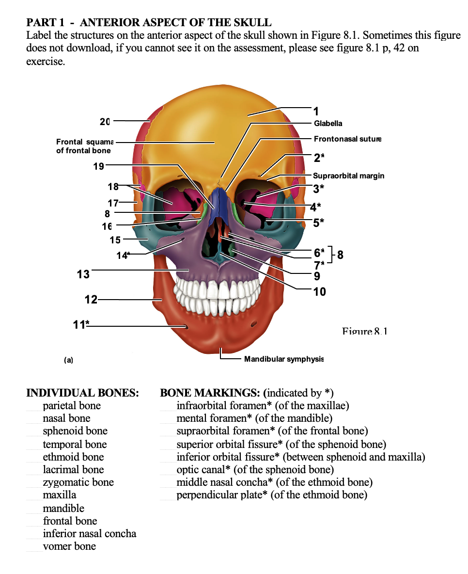
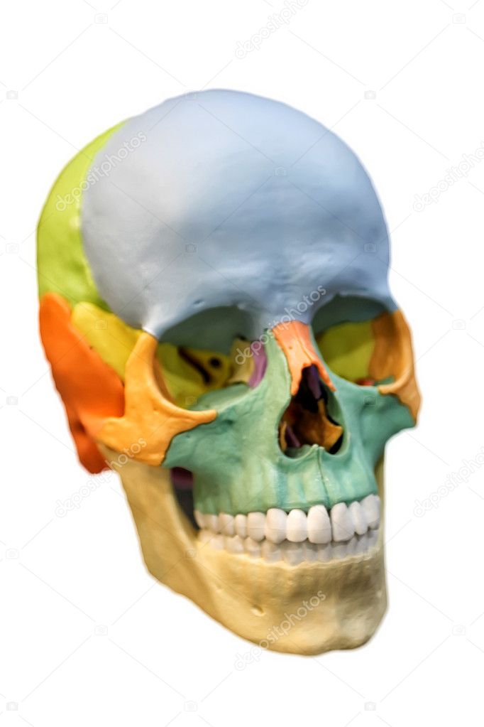
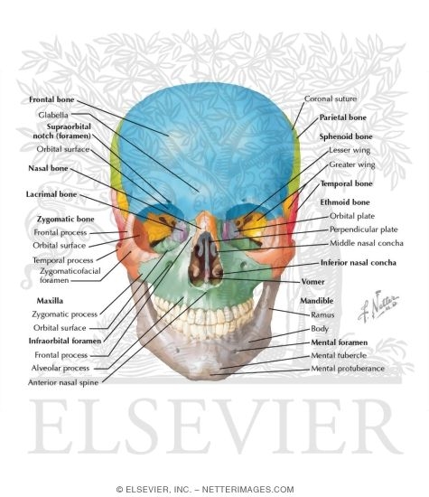
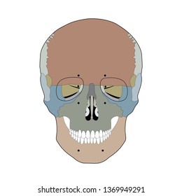
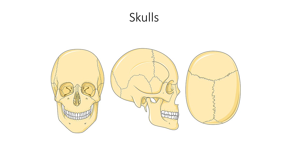
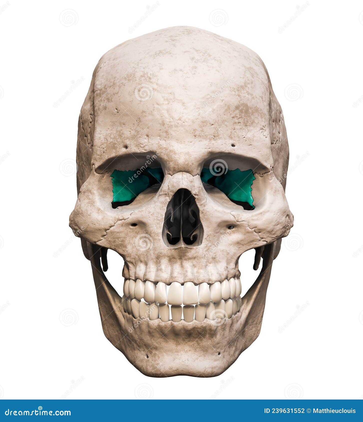
45 anterior view of skull unlabeled
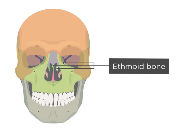
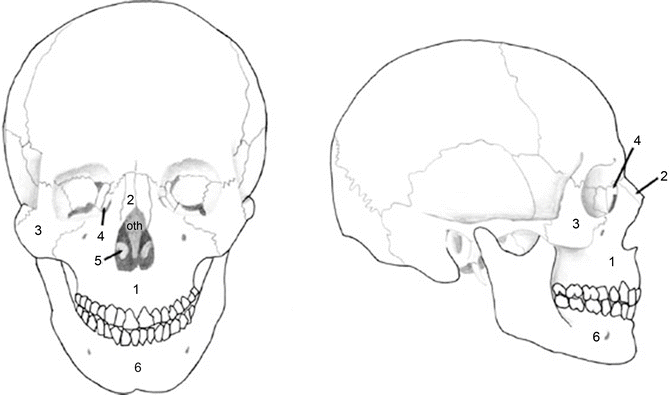
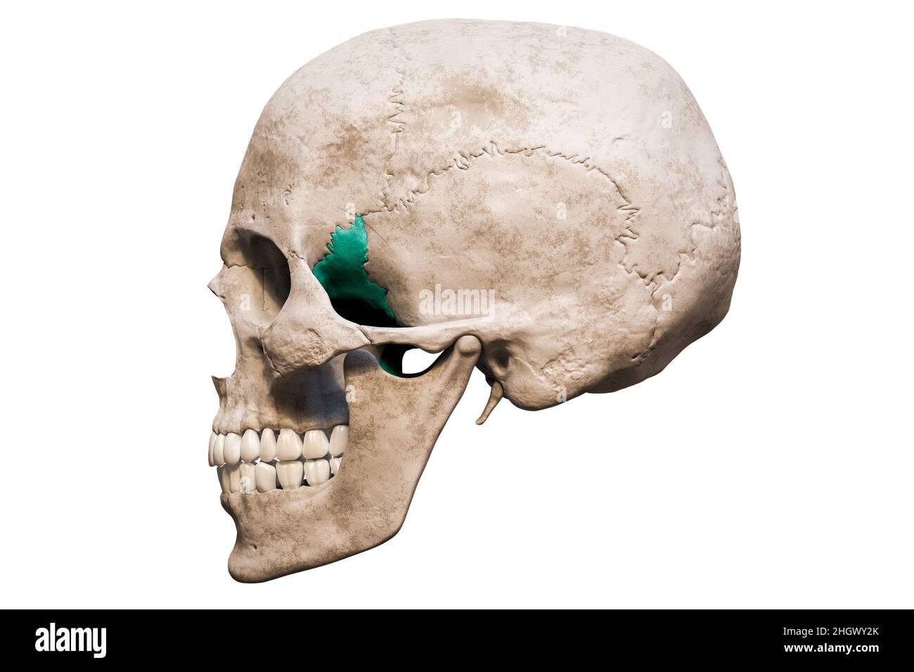
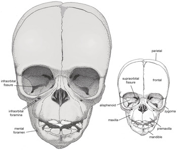
Skull anatomy: Anterior and lateral views of the skull | Kenhub The bones of the skull that are visible from an anterior and a lateral view are the following: the sphenoid bone (with the greater and the lesser wings) the frontal bone (especially the orbital surface) the zygomatic bone the maxilla the mandible the nasal bones the ethmoid bones the parietal bone and the temporal bone Bones of the Foot - Tarsals - Metatarsals - TeachMeAnatomy Tarsals - a set of seven irregularly shaped bones. They are situated proximally in the foot in the ankle area. Metatarsals - connect the phalanges to the tarsals. There are five in number - one for each digit. Phalanges - the bones of the toes. Each toe has three phalanges - proximal, intermediate, and distal (except the big toe ...
Male And Female Reproductive System Quiz! - ProProfs 1. A muscular tube that passes upward alongside the testes and transports semen. A sac-like structure attached to the vas deferens and to the side of the bladder. Controls temperature of testes. Reduces temperature of testes. 2. When semen is pushed through and out of a male's body through the urethra is a process called_______.

Anterior view of skull unlabeled
Skull Anatomy Labeling - Human Anatomy - GUWS Medical Label the anterior bones and features of the skull. (If the line lacks the word bone, label the particular feature of that bone.) middle nasal concha ethmoidal sinuses crista galli 4. Complete Parts A and B of Laboratory Report 13. 5. Examine the facial bones of the articulated and sectioned skulls and the corresponding disarticulated bones. NOWinBRAIN: a Large, Systematic, and Extendable Repository of 3D ... Despite the tremendous development of various brain-related resources, a large, systematic, comprehensive, extendable, and beautiful repository of 3D reconstructed images of a living human brain expanded to the head and neck is not yet available. I have created such a novel repository and populated it with images derived from a 3D atlas constructed from 3/7 Tesla MRI and high-resolution CT ... Sternum - Anatomy, Parts, Location, Functions, & Diagram Sternum, commonly called breastbone, is a long, flat bone located in the midline of the chest. The word 'sternum' has been derived from the ancient Greek word ' sternon ', meaning 'chest'. The bone covers and protects the thoracic organs, such as heart, and lungs from any external shock. Where is the Sternum Bone Located
Anterior view of skull unlabeled. Dog Anatomy from Head to Tail - dummies Head's up on dog parts. Starting from the head, a dog is made up of the. Nose: Dog noses are often cold and wet, and of course, they usually get stuck where they're not wanted. The muzzle (foreface) comprised of the upper and lower jaws. The stop is an indentation (sometimes nonexistent) between the muzzle and the braincase or forehead. Labeled imaging anatomy cases | Radiology Reference Article ... This article lists a series of labeled imaging anatomy cases by body region and modality. Brain CT head: non-contrast axial CT head: non-contrast coronal CT head: non-contrast sagittal CT head: angiogram axial CT head: angiogram coronal CT... Anatomical Position: Body Planes and Sections - EZmed The transverse plane is the only horizontal plane, and it divides the body into top (superior) and bottom (inferior) sections. An easy trick to remember the transverse plane is to again use the name. The "Transverse" plane will give you a "Top View" of the body as it divides the body into upper and lower portions. Inferior view of the base of the skull: Anatomy | Kenhub This foramen is also known as the anterior palatine foramina, and can be found in the anterior most section of the midline of the maxillary bone. They allow the nasopalatine nerves and the sphenopalatine artery to enter from the floor of the nasal cavity. Forming the roof of the nasopharynx are the choanae superior to the hard palate.
Lateral View Of The Skull Worksheet Answers - Google Groups Pictures of skulls that are unlabeled and remains empty boxes for students to add labels Skull is pictured from many angles. Detailed information between skull lateral views as a worksheet answers... Horse Skeleton Anatomy - Osteological Features of Bones from Equine ... The skull of a horse is long and four-sided. You will find an extensive foramen lacerum in the horse skull. There is no cornual process in horse skull. The fusion between the two haves of the mandible is complete. There is no acromion process in the scapula of a horse. The glenoid notch is distinct and deep. NOWinBRAIN 3D neuroimage repository: Exploring the human brain via ... A view image sequence contains five images. An appearance image sequence typically contains three images (non-parcellated unlabeled, parcellated unlabeled, and parcellated labeled); its number is seven when all three cortical color maps are employed (and it may be even higher when additionally two white matter color maps are considered). Anatomical Terms & Meaning: Anatomy Regions, Planes, Areas, Directions The ventral cavity is on the front (anterior) of the body and is divided into the thoracic (chest) and abdominopelvic cavities. Dorsal Cavity The dorsal cavity is further divided into the following subcavities: The cranial cavity contains the brain The spinal (or vertebral cavity) contains the spinal cord. Ventral Cavity
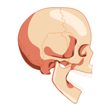
Scapula - Parts, Anatomy, Location, Functions, & Labeled Diagram It is the anterior surface of the scapula facing the thoracic cage or ribcage. It has a large concave depression over most of the surface, called the subscapular fossa, from where the rotator cuff muscle subscapularis originates. This region is marked by some longitudinal ridges, out of which a thick ridge joins the lateral border. Blank Skull Bones Cranial - skull diagram superior view of floor of ... Blank Skull Bones Cranial. Here are a number of highest rated Blank Skull Bones Cranial pictures on internet. We identified it from trustworthy source. Its submitted by admin in the best field. We say you will this kind of Blank Skull Bones Cranial graphic could possibly be the most trending subject later than we part it in google benefit or ... Anatomy, Back, Spinal Meninges - StatPearls - NCBI Bookshelf The spinal cord and brain are encased within three layers of tissue called the meninges. The spinal meninges specifically enclose the spinal cord and stretch from the brainstem down to the filum terminale. The layers of the meninges are, from deep to superficial, the pia mater, the arachnoid mater, and the dura mater. The names of these layers give information on their qualities. Pia, which is ... Skull (lateral view) | Radiology Reference Article - Radiopaedia the beam travels laterally, with 0° of angulation, through a point ~4 cm above the external auditory meatus collimation superiorly to include skin margins inferiorly to include base of skull anteriorly to include frontal bone posteriorly to the skin margins orientation landscape detector size 24 cm x 30 cm exposure 60-70 kVp 10-20 mAs SID 100 cm
Body Cavities and Membranes: Labeled Diagram, Definitions We are looking at a side view of the brain and skull below called a sagittal view. As mentioned before, the cranial cavity is enclosed by the cranium or skull which is highlighted in green. You can see how the skull has formed an empty space or cavity. This space created by the skull is called the cranial cavity which is shown in yellow.
Anterior view of skull Quiz - PurposeGames.com This is an online quiz called Anterior view of skull. There is a printable worksheet available for download here so you can take the quiz with pen and paper. Your Skills & Rank. Total Points. 0. Get started! Today's Rank--0. Today 's Points. One of us! Game Points. 25. You need to get 100% to score the 25 points available.
Bones of the Foot | Tarsal bones - Metatarsal bone - Geeky Medics Figure1.Tarsal bones of the foot (superior view) 1 Talus. Talus (Latin for ankle) talus is the most superior bone of the tarsus and rests on top of the calcaneus. Three areas of articulation form the ankle joint: . T he superomedial aspect of the talus articulates with the medial malleolus of the tibia; T he superior aspect of the talus articulates with the distal portion of the tibia
Bones of the Skull | Skull Osteology | Anatomy | Geeky Medics The calvarium, also known as the roof or skull cap, consists of three bones: Frontal bones. Parietal bones. Occipital bones. These bones protect the brain superiorly, but also provide an anchor for important muscles of facial expression and eye movement. The parts of these bones that lie inferior to the brain are considered to be a part of the ...
Anatomical diagrams of the brain - e-Anatomy - IMAIOS The study of the arterial supply of blood to the brain is facilitated by a diagram showing the cerebral arterial vascular areas in lateral and medial views and axial and coronal section and by diagrams of arteries forming the Willis' circle (internal and vertebral carotid arteries, basilar artery, anterior and posterior communicating arteries ...
Brain | Radiology Reference Article | Radiopaedia.org The brain is the vital neurological organ composed of: cerebrum diencephalon brainstem midbrain pons medulla cerebellum The brain is housed in the neurocranium of the skull and bathed in cerebrospinal fluid. It is continuous with the cerv...
Anatomy of the cranial nerves - IMAIOS This human anatomy module is about the cranial nerves. It consists of 15 vector anatomical drawings with 280 anatomical structures labeled. It is intended for the use of medical students working on human anatomy, student nurses, physiotherapists, electro-radiological technicians and residents - especially those working in neurology, neurosurgery, otolaryngology - and for any physician ...
Skull Frontal View - lateral and frontal view of normal skull ... Skull Frontal View. Here are a number of highest rated Skull Frontal View pictures upon internet. We identified it from honorable source. Its submitted by dispensation in the best field. We agree to this kind of Skull Frontal View graphic could possibly be the most trending subject subsequently we part it in google benefit or facebook.
Axial Skeleton Labeling Worksheet - groups.google.com Bones Of one Skull Anterior View Worksheet Answers. Label the 4 parts of the vertebrae below color the tailbone 05. Identify specifically each search the vertebra types shown in the diagrams below....
Occipital Bone: Anatomy, Function, and Treatment - Verywell Health As a person ages, their occipital bones will fuse to the other bones of their skull. Your sphenoid bone, which is located in the middle of your skull, will fuse with the occipital bone between the ages of 18 and 25. Then, between the ages of 26 and 40, the parietal bones at the top of your head and occipital bone will fuse together.
Sternum - Anatomy, Parts, Location, Functions, & Diagram Sternum, commonly called breastbone, is a long, flat bone located in the midline of the chest. The word 'sternum' has been derived from the ancient Greek word ' sternon ', meaning 'chest'. The bone covers and protects the thoracic organs, such as heart, and lungs from any external shock. Where is the Sternum Bone Located
NOWinBRAIN: a Large, Systematic, and Extendable Repository of 3D ... Despite the tremendous development of various brain-related resources, a large, systematic, comprehensive, extendable, and beautiful repository of 3D reconstructed images of a living human brain expanded to the head and neck is not yet available. I have created such a novel repository and populated it with images derived from a 3D atlas constructed from 3/7 Tesla MRI and high-resolution CT ...
Skull Anatomy Labeling - Human Anatomy - GUWS Medical Label the anterior bones and features of the skull. (If the line lacks the word bone, label the particular feature of that bone.) middle nasal concha ethmoidal sinuses crista galli 4. Complete Parts A and B of Laboratory Report 13. 5. Examine the facial bones of the articulated and sectioned skulls and the corresponding disarticulated bones.





:background_color(FFFFFF):format(jpeg)/images/library/7569/Inferior_nuchal_line_.png)

:background_color(FFFFFF):format(jpeg)/images/library/11336/labaled_diagram_main_bones_of_skull.jpg)

:background_color(FFFFFF):format(jpeg)/images/library/623/os_sphenoidale_large_BW4XMAi2VQjx0tdO4RVgcw.png)








:watermark(/images/watermark_only_sm.png,0,0,0):watermark(/images/logo_url_sm.png,-10,-10,0):format(jpeg)/images/anatomy_term/skull/eEsfu70EOMx1TlBf5tYAiA_Go0bFvBvzClwSivuaiELg_head_01.png)
Post a Comment for "45 anterior view of skull unlabeled"